A digital X-ray, or digital radiography, is a modern type of X-ray that utilises digital sensors instead of photographic film, as with a traditional X-ray. The image captured is converted to digital data immediately and is available for review within seconds.
The digital X-ray procedure is highly similar to traditional X-rays; it is the technology that is very different. Bursts of radiation still pass through the body and form an image based on how much radiation passes through the different organs, but rather than using photographic film to capture the image, digital sensors are used.
Digital X-ray sensors usually come in the form of active matrix flat panels made up of a detection layer over an active matrix array of thin film transistors and photodiodes. These sensors are able to convert the image into digital form in real time, allowing the doctor to view the results on a computer immediately.
Digital X-rays, much like traditional X-rays, allow the doctor to examine inside a patient’s body. This may be useful to observe the extent of the damage done during an injury, including bone breaks and fractures. They can also detect masses in soft tissue, which could lead to the discovery of a tumour or other illnesses.
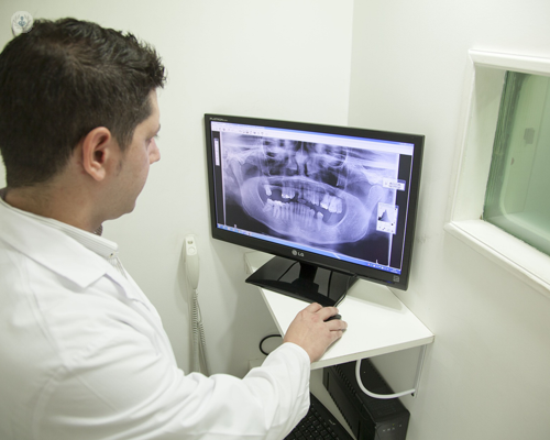
Digital X-rays are of particular interest in dentistry, where the rapid availability of the results means that a dentist can enhance the images by controlling the exposure in real time and therefore can obtain clear and detailed results that can be shared with the patient immediately. The clarity of digital X-rays makes them superior to traditional X-rays in terms of finding tiny fractures and imperfections in the teeth.
Digital X-rays have several key advantages over other options: